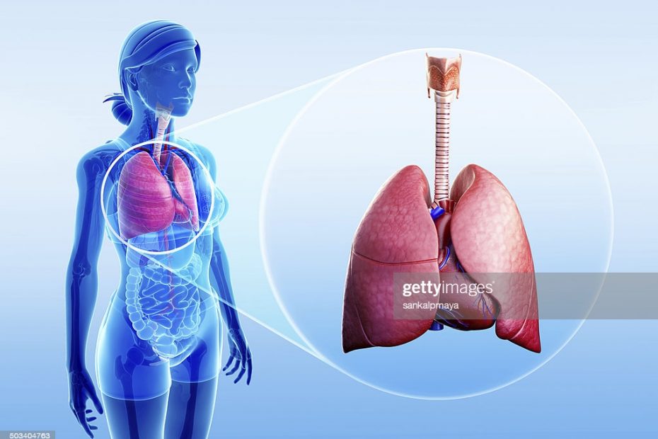
Empyema is a disease in which the area between the lungs and the inner surface of the chest wall contains pus. This Field is referred to as pleural room or space. Pus is a fluid that contains immune cells, bacteria, and dead cells. Pus cannot be coughed up from the pleural space. Instead a needle or surgery is required to drain it. Pyothorax or purulent pleuritis is also called empyema. Empyema normally occurs after pneumonia, which is an infection of lung tissue.
CAUSES
After you have pneumonia, empyema may develop. Pneumonia can be caused by several different kinds of bacteria, but two common causes are streptococcus pneumonia and staphylococcus aureus. Empyema can sometimes occur after you’ve had chest surgery. The pleural cavity can be contaminated with bacteria during surgery.
Naturally, the pleural space has some fluid, but infection can cause fluid to build up more quickly than it can be absorbed. The fluid is then contaminated with the bacteria and subsequently white blood cells and dead cells which thickens the fluid. It can cause the lining of your chest cavity and lungs to stick together and form pockets. This is known as Empyema. The lungs will not be able to fully inflate, which may contribute to trouble breathing.
CONDITIONS THAT CAN PLACE YOU AT RISK!
Bacteria pneumonia is the greatest risk factor for Empyema. Empyema is most common in children and older adults. Overall, it is fairly rare however occurring in less than 1% of children with pneumonia in a study.
Chances of Empyema after pneumonia can also be increased by having the following conditions;
Bronchiectasis, chronic obstructive pulmonary disease { COPD }, alcoholism, Diabetes and Recent trauma or surgery.
SYMPTOMS
Empyema can be straightforward or complex
Simple or straightforward Empyema
In the early phase of the disease, basic Empyema occurs. If the pus is free flowing, an entity has this shape. The signs of this type of Empyema include;
Shortness of breath, dry cough with fever, sweating, chest pain that may be identified as stabbing when breathing, headaches and loss of appetite.
Complex Empyema
In the later stages of the disorder, complex Empyema occurs. The inflammation in complex empyema is more severe. Scar tissue can form and break the cavity of the chest into smaller cavities. This is called loculation, and this is harder to treat.
Other complex Empyema symptoms are;
Breathing problems, decreased breath sounds, weight loss and chest pain.
Complications of Empyema
These include lung collapse and sepsis. Sepsis signs include;
Fever, rapid respiration, increased heart rate and Low blood pressure
Sudden sharp chest pain and shortness of breath that gets worse when coughing or breathing can be caused by collapsed lung.
Management
Management involves draining the pus and fluid from the pleura and antibiotics to treat the infection. The exact type of antibiotic depends on the type of bacteria.
The technique used to drain the pus relies on the Empyema process. In simple empyema, it is possible to insert a needle into the pleural region to remove the fluid. This is known as percutaneous thoracentesis. Complex empyema on the other hand typically requires a surgical procedure under anesthesia to remove the pus.
Photo credit :
Breathe.ersjournals.com

Pharm. Fatima Ajide J. is a current pharmacology student at Olabisi Onabanjo University.
She has a passion in researching about the interaction between organs and drugs (i. e. the Pharmacodynamic and Pharmacokinetics).
A practicing Muslim and hobbies are tourism, reading and Research.