READERS’/FANS’ CORNER
Ewing sarcoma is a cancerous tumour which occurs in the bone or other body tissue like cartilage or nerves. Broadly speaking, there are three types of Ewing sarcoma which are: Ewing sarcoma of bone, extraosseous ewing sarcoma, Askin tumour (Ewing sarcoma involving the chest wall), peripheral primitive neuroectodermal tumour.
About 90% of Ewing Sarcoma are Ewing sarcoma of the bones. Bones often involved are thigh bone, chest bone, pelvis or shoulder blade.
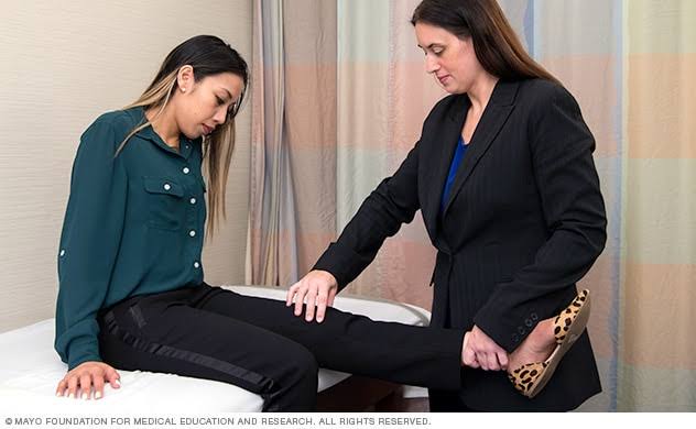
Ewing sarcoma usually occur in children and young adults. It is the second most common bone cancer in children. Osteosarcoma being first on the list. Affected individual often complained of bone stiffness, pain, swelling, tenderness of the bone or surrounding tissue. Often, children have fever that does not go away. Ewing sarcoma can cause weakening of the involved bone, and affected individuals may have a broken bone with no obvious cause. Other symptoms are: anorexia and weight loss. Compare Ewing sarcoma with Osteopetrosis. https://www.healthgist.net/health-gist-with-just-a-minor-fall-her-bone-breaks-the-same-happened-to-her-mother-osteopetrosis/
It is not uncommon for Ewing Sarcoma to spread to other part of the body usually lungs or other bone.
DOCTORS’ CORNER
The most common mutation that causes Ewing sarcoma involves two genes, the EWSR1 gene on chromosome 22 and the FLI1 gene on chromosome 11. In many patients, the first presenting complaint is intermittent locoregional pain that does not disappear at night. As the tumour grows, palpable mass is seen at the affected site.
INVESTIGATIONS
Plain radiograph: tumour is seen within the diaphyseal region with triad of Codman’s triangle, osteolysis and calcified spicules within the tumour mass. Codman’s triangle is seen when an aggressive bone lesion grows faster than the overlying periosteum. It is also seen in osteosarcoma and osteomyelitis. https://www.healthgist.net/osteomyelitis-a-cause-of-bone-pain/
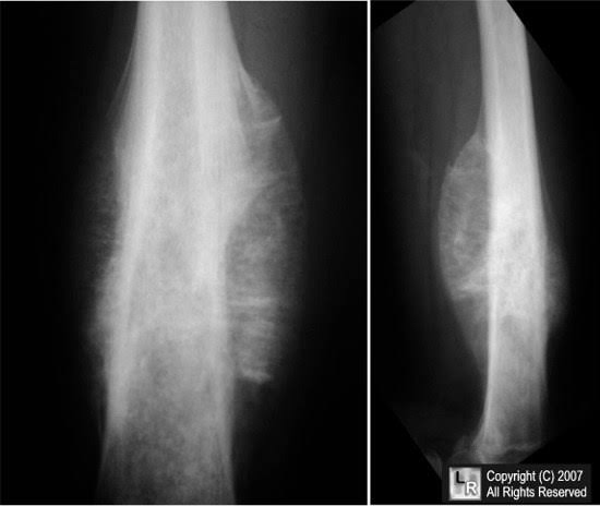
Image credit: google
MRI of the region: It shows exact site, local spread and size of the tumour. Computed tomography helps reveal possible metastasis to the thorax.
Biopsy: This is diagnostic using open biopsy and NEVER fine-needle because of risk of tumour seeding.
Positron Emission Tomography helps to detect possible skeletal metastasis.
Routine laboratory investigation may reveal elevated erythrocyte sedimentation rate, leukocytosis and elevate serum lactate dehydrogenase.
TREATMENT
Treatment lasts 6-9months and consists of alternating courses of 2 chemotherapy regimen: (1) Vincristine, doxorubicin and cyclophosphamide and (2) ifosfamide and etoposide.
Management of tumour site is very critical in Ewing sarcoma. This might involve limb salvage with extensive margins or amputation.
CREDIT:
U.S. National Library of Medicine
Medline

Dr. Adeyemo Olusola is a medical graduate of Olabisi Onabanjo University, Ogun State, Nigeria along with certificate in advanced diploma in Principles of Nutrition, Management and Leadership, Dublin and Certificate in Global Health from London School of Hygiene and Tropical Medicine. In addition to his numerous certifications, he is a certified Telemedicine Physician from Harvard Medical School, USA. He is an avid reader of books from different oases of life, expert in data analysis. “So many a time, I have seen people die avoidable death because of lack of knowledge or information, falling victim of fate. There is then a necessity laid on us to help arm our society to the teeth, as a healthy society cannot be detached from an informed one. Hence, there is need for healthgist.net. We hope you will have a wonderful stay on our website.”