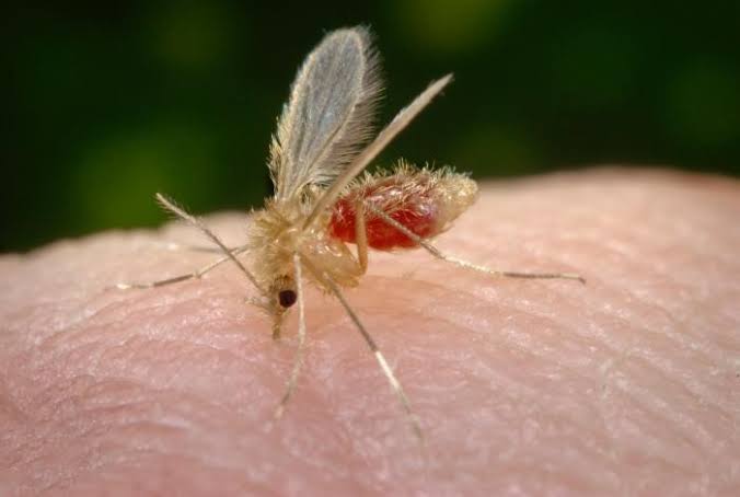
The parasites that cause Leishmaniasis belong to the leishmania genus. There are more than 20 pathogenic species. Female sand-flies transmit the parasites by biting and feeding on humans, similarly to mosquitoes. The sand-fly becomes infected when taking blood from reservoir hosts.
WORLD STATISTICS
Leishmaniasis is endemic in 88 countries throughout Latin America, Africa, Asia, and Southern Europe. 350 million people worldwide are thought to be at risk. Around 12 million people worldwide are reported to be infected with the parasites with about 1.5 million new cases of cutaneous Leishmaniasis annually.
90% of cutaneous Leishmaniasis occurs in Afghanistan, Iran, Saudi Arabia, Syria, Brazil and Peru, 90% of all visceral Leishmaniasis occurs in Bangladesh, Brazil, India, and the Sudan. Likewise, 90% of muco-cutaneous Leishmaniasis occurs in Bolivia, Brazil and Peru.
Children are more affected than adults and incubation periods (period from infection to the onset of clinical signs) can vary from a few days to many months.
Leishmaniasis is a parasitic disease capable of causing a spectrum of clinical presentations ranging from cutaneous ulcerations (open sores) to systemic infections (generalized infection).
THE SPECTRUM OF LEISHMANIASIS
CUTANEOUS LEISHMANIASIS – A localized infection of the skin which is caused by leishmania tropica
MUCOCUTANEOUS LEISHMANIASIS- Localized infection of muco-cutaneous junctions and mucosal surfaces.
VISCERAL LEISHMANIASIS- Characterized by irregular fever, liver and spleen enlargement, lymph node enlargements, anemia etc. It is caused by leishmania donovani.
CLINICAL STATUS
Leishmaniasis can be sub-clinical with no obvious signs. It can also present with disseminated infections which can lead to death.
The first sign is usually a small erythematous papule that may appear immediately following a bite from a sand fly but usually appears 2 to 4 weeks later.
The papule slowly enlarges over a period of several weeks and assumes a more dusky purplish hue. The lesion eventually becomes crusted in the center.
When the crust is removed, there is a shallow ulcer, often with a raised and indurated border.
The ulcer is typically painless. Small papules that enlarge and ulcerate at the center produce a volcano-shaped wet lesion.
Lesions can be typical, usually a painless, sharply delineated ulcer surrounded by a purplish raised border. Dry lesions with smooth or hyperkeratotic surfaces can also occur.
LEISHMANIASIS IN HIV PATIENTS
Co-infection with HIV can amplify the immune defect and increase disease severity and morbidity with local dissemination (caused by the spread of either parasites or their antigens away from the site of a primary lesion) occurring commonly.
This may be the first manifestation leading to the diagnosis of HIV infection. Human immunodeficiency virus co-infection with cutaneous Leishmaniasis may be associated with unusually severe presentations, higher rates of recurrence and re-infection, and lower cure rates with established treatments. Disseminated cutaneous Leishmaniasis is clinically characterized by multiple, atypical, widespread infiltrations of skin lesions.
PREVENTION
Covering of the whole body to avoid bites from sand-flies,
Sleeping under treated mosquito nets and
Using insecticides in sleeping and living areas.
DIAGNOSIS
1. THE LEISHMANIN SKIN TEST (LST) (Montenegro test)
In cutaneous Leishmaniasis the leishmanin skin test will become positive 2-3 months after the appearance of the lesion and may remain positive for life.
2. DIRECT EXAM
Microscopy of tissue fluid from aspiration, scrapings, or biopsy smears can be performed after staining with Giemsa stain.
3. CULTURE
Culture of aspirates, scrapings, and fresh skin biopsies.
4. SEROLOGIC TECHNIQUES
The indirect methods detect anti-Leishmania antibodies, they include, the direct agglutination, Immuno-fluorescence and Immuno-blot tests.
TREATMENT
Localized cutaneous lesions Cryotherapy, Thermotherapy, Curettage, Photodynamic Therapy, Amphotericin, paromomycin and antimonials are recommended medications.
Photo credit: Jove.com, Journals.rcni.com and Medicine net.com

Pharm. Fatima Ajide J. is a current pharmacology student at Olabisi Onabanjo University.
She has a passion in researching about the interaction between organs and drugs (i. e. the Pharmacodynamic and Pharmacokinetics).
A practicing Muslim and hobbies are tourism, reading and Research.